
This will be my hearing system. Information from the Cochlear UK website.
Royal National Throat, Nose and Ear Hospital
Surgeon Mr Azhar Shaida Cochlear Implant Department
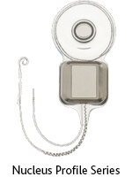
Nucleus® Implant portfolio
A lifetime of reliable hearing.
Cochlear’s range of Nucleus Implants is
the result of more than 30 years of ground breaking, collaborative
clinical and technical research work.
Our scientists and engineers understand that we all have very different anatomy. That’s why they’ve designed the widest range of implants and electrodes to deliver the best possible sound resolution, coverage and comfort for your unique ear.
Our scientists and engineers understand that we all have very different anatomy. That’s why they’ve designed the widest range of implants and electrodes to deliver the best possible sound resolution, coverage and comfort for your unique ear.
Implants designed for your ear
You and your surgeon can now choose from two Nucleus Implant Series, each offering sophisticated electronics, innovative designs and quality materials.The thinnest implant in the world
Our latest generation of implants - the Nucleus Profile Series - is the thinnest implant in the world, designed to better conform to the natural shape of the head, making it more discreet to wear.A state-of-the art manufacturing facility has been established specially for the Nucleus Profile Series and all of our future generations of implants.

Electrodes to suit your hearing needs
The precurved design of the Contour Advance Electrode allows the surgeon to better position the electrode array in very close proximity to your hearing nerve for more focused and accurate stimulation. The world’s thinnest full length Slim Straight electrode provides excellent electrical stimulation outcomes, with proven preservation of residual hearing.Implant Design
The inner ear, or cochlea, is a small and
delicate spiral-shaped structure. Tiny hair cells within the cochlea
convert sound into signals, which are then sent to the brain via the
hearing nerve.
Hearing with a cochlear implant is different to normal hearing.
Normal hearing works by sending sound vibrations to hair cells within the cochlea. A cochlear implant uses an electrical impulse to stimulate a different set of cells called the spiral ganglion cells. The spiral ganglion cells are located in a different area of the cochlea and connected to the hair cells.
Spiral ganglion cells are mostly concentrated in an area that we call the ‘hearing zone’. This area is the most responsive to electrical stimulation from a cochlear implant. The hearing zone does not extend deep into the cochlea.
As such, Cochlear’s philosophy is to deliver electric stimulation to the spiral ganglion cells located in the hearing zone to achieve optimal hearing performance, and to avoid deeper insertion in order to reduce the risk of apical stimulation or insertion trauma. This philosophy underpins the development of our range of electrodes.


Electrode design and hearing performance
With pre-curved, perimodiolar electrodes the stimulation contacts are placed closer to the spiral ganglion cells through a design that matches the natural shape of the cochlea. After insertion, the electrode array sits in a relaxed resting position without applying any force to either the lateral or the modiolar wall. Research has shown that perimodiolar electrodes deliver unprecedented hearing performance, a more focused stimulation and greater power efficiency.Lateral wall electrodes have also been shown to deliver excellent hearing performance in both electric-only and combined electro-acoustic stimulation modes.
Special design features such as a basal stiffener, Softip™ smooth lateral wall surface or handle give the surgeon control over insertion depth and therefore the ability to achieve optimal coverage of the hearing zone in varying cochlear sizes.
Basal strength provides control and tactile feedback for the surgeon to increase the predictability of insertion and minimise buckling, which is often associated with significant insertion trauma. Apical flexibility is important to minimise insertion forces which may cause trauma to delicate lateral wall structures.
The design and shape of the electrode tip needs to protect the delicate internal structures of the cochlea and therefore varies in shape of perimodiolar and lateral wall electrodes.
Half-band electrode contacts featured on all of our electrodes ensure there is a smooth silicone surface facing the lateral wall to minimise friction trauma during insertion.
 |
| My implant in position |
MRI compatibility
MRI (Magnetic Resonance Imaging) is a
common scan used for medical imaging procedures. MRI uses a very
powerful magnet to provide detailed images of a person’s internal organs
and tissue. It is often used to provide early detection of many
different conditions so that treatment can be more effective and timely.
As implanted medical devices can interfere with MRI scans, it’s important to consider the compatibility of this increasingly popular technology with your choice of cochlear implant.
As implanted medical devices can interfere with MRI scans, it’s important to consider the compatibility of this increasingly popular technology with your choice of cochlear implant.
How does a cochlear implant affect MRI?
The internal implant contains a magnet, which holds the external sound processor coil in place. When placed in an MRI scanner, this magnet can cause a blur or ‘artifact’ over the medical image, which may hinder the doctor’s ability to make an accurate diagnosis of brain scans. As a quarter of all MRI scans are performed on the brain, having the flexibility to remove the internal magnet if required is an important consideration when choosing a cochlear implant.Other brands may not feature a removable magnet
This means that recipients may be limited to older MRI technology or may need their entire implant including the electrode removed in surgery, even for a scan of a different part of the body such as the knee.Implant removal requires surgery. Once an implant is removed, it cannot be re-used, meaning a new implant is required and therefore further surgery.
No comments:
Post a Comment